Conjoint Tendon Shoulder Anatomy - Schematic Representation Of The Right Shoulder Anterior View The Download Scientific Diagram. The long head of biceps (lhb) is a very important tendon that travels through the shoulder joint (glenohumeral joint). If you tear your biceps tendon at the shoulder, you may lose some strength in your arm and have pain when you forcefully turn your arm from palm down to palm up. Learn vocabulary, terms and more with flashcards, games and other study tools. The shoulder joint is the connection between the chest and the upper extremity. Anterior projection conjoint tendon laterjet impingement.
The conjoint tendon (previously known as the inguinal aponeurotic falx) is a structure formed from the lower part of the common aponeurosis of the internal oblique muscle and the transversus abdominis as it inserts into the crest of the pubis and pectineal line immediately behind the superficial inguinal ring. It gets its name from the fact that it is often continuous or conjoined with the tendon of the internal oblique, another of the abdominal muscles. In this episode of eorthopodtv, orthopaedic surgeon randale c. The long head of biceps (lhb) is a very important tendon that travels through the shoulder joint (glenohumeral joint). Robin smithuis and henk jan van der woude.
Seminar On Applied Anatomy And Surgical Approaches To Shoulder from image.slidesharecdn.com The shoulder joint is formed the rotator cuff is a collection of muscles and tendons that surround the shoulder, giving it. This image shows the anatomy of the shoulder joint from posterior view displaying the bones, tendons and muscles of the joint in shoulder joint:anatomy,movement & muscle involvement. Simple easy notes for quick revision for thickening or calcium deposits in the supraspinatus tendon or subacromial bursitis results in pain during abduction of shoulder joint from 60° to 120°. Knowledge of the shoulder will help you understand the different shoulder problems. Anatomy of the canine shoulder (scapula, humerus, ligaments, shoulder joint, muscles and tendons) on ct. Robin smithuis and henk jan van der woude. The shoulder joint is the connection between the chest and the upper extremity. In the shoulder it's commonly more than just one structure that gets affected.
Knowledge of the shoulder will help you understand the different shoulder problems.
Normal anatomy and pathology on mri. Normal anatomy, variants and checklist. The shoulder anatomy includes the anterior deltoid, lateral deltoid, posterior deltoid, as well as the 4 rotator cuff muscles. Related online courses on physioplus. If you tear your biceps tendon at the shoulder, you may lose some strength in your arm and have pain when you forcefully turn your arm from palm down to palm up. Upper limb trauma programme injuries. It gets its name from the fact that it is often continuous or conjoined with the tendon of the internal oblique, another of the abdominal muscles. Knowledge of the shoulder will help you understand the different shoulder problems. Anterior graphic of the shoulder. In this episode of eorthopodtv, orthopaedic surgeon randale c. The conjoint tendon (previously known as the inguinal aponeurotic falx) is a structure formed from the lower part of the common aponeurosis of the internal oblique muscle and the transversus abdominis as it inserts into the crest of the pubis and pectineal line immediately behind the superficial inguinal ring. Shoulder muscles and shoulder tendons. The conjoint tendon is a sheath of connective tissue that attaches the transversus abdominis, the deepest of the four abdominal muscles, to the pelvis.
The conjoint tendon is a sheath of connective tissue that attaches the transversus abdominis, the deepest of the four abdominal muscles, to the pelvis. The interfoveolar ligament, seen from in front. Revision anterior stabilization of the shoulder presents a challenge to the surgeon and carries a higher risk of recurrent dislocation than primary repair. The biceps muscle has two tendons at the shoulder, called the long head and short head. This image shows the anatomy of the shoulder joint from posterior view displaying the bones, tendons and muscles of the joint in shoulder joint:anatomy,movement & muscle involvement.
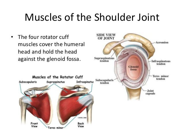
The Shoulder Musculoskeletal Key from musculoskeletalkey.com Professor of radiology and orthopaedic surgery department of radiology university of california san francisco. Robin smithuis and henk jan van der woude. The subscapularis, teres minor, supraspinatus, and infraspinatus. Muscles allow us to move by pulling on bones. Four muscles make up the rotator cuff: Upper limb trauma programme injuries. The conjoint tendon, also known as henle's ligament, forms when the medial fibers of the internal oblique aponeurosis unite with the deeper fibers of the transversus abdominis aponeurosis. Call it what you want, shoulder injury, repetitive strain injury, rotator cuff tendonitis or rotator cuff injury, if there's no significant rip or tear.
If you tear your biceps tendon at the shoulder, you may lose some strength in your arm and have pain when you forcefully turn your arm from palm down to palm up.
The subacromial bursa lies on the top portion of the supraspinatus tendon. Sechrest, md narrates an animated tutorial on the basic anatomy of the shoulder. These are the main ligaments that help to stabilize the joints of. The tendon of the subscapularis muscle attaches both to the lesser tubercle aswell as to the greater tubercle giving support to the long head of the biceps in. Your biceps tendons attach the biceps muscle to bones in your shoulder and in your elbow. Injuries to the rotator cuff are common, but. It gets its name from the fact that it is often continuous or conjoined with the tendon of the internal oblique, another of the abdominal muscles. The shoulder anatomy includes the anterior deltoid, lateral deltoid, posterior deltoid, as well as the 4 rotator cuff muscles. Biceps as the name implies originates from two points. Four muscles make up the rotator cuff: The name gets its origin from its structure which is often conjoined or continuous with. Ligaments are soft tissue structures that connect bones to bones. Muscles allow us to move by pulling on bones.
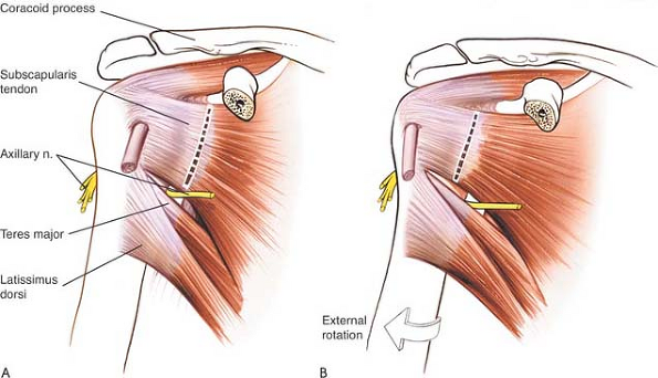
Learn vocabulary, terms and more with flashcards, games and other study tools. One tendon might have it worse, but it's never isolated to just one tendon. Knowledge of the shoulder will help you understand the different shoulder problems. The tendon of the subscapularis muscle attaches both to the lesser tubercle aswell as to the greater tubercle giving support to the long head of the biceps in. There are several important ligaments in the shoulder.
Shoulder Anatomy And Techniques Radiology Key from radiologykey.com Anatomy of the canine shoulder (scapula, humerus, ligaments, shoulder joint, muscles and tendons) on ct. Learn vocabulary, terms and more with flashcards, games and other study tools. The conjoint tendon (previously known as the inguinal aponeurotic falx) is a structure formed from the lower part of the common aponeurosis of the internal oblique muscle and the transversus abdominis as it inserts into the crest of the pubis and pectineal line immediately behind the superficial inguinal ring. This image shows the anatomy of the shoulder joint from posterior view displaying the bones, tendons and muscles of the joint in shoulder joint:anatomy,movement & muscle involvement. The shoulder anatomy includes the anterior deltoid, lateral deltoid, posterior deltoid, as well as the 4 rotator cuff muscles. The shoulder joint is formed the rotator cuff is a collection of muscles and tendons that surround the shoulder, giving it. If you tear your biceps tendon at the shoulder, you may lose some strength in your arm and have pain when you forcefully turn your arm from palm down to palm up. Revision anterior stabilization of the shoulder presents a challenge to the surgeon and carries a higher risk of recurrent dislocation than primary repair.
Anatomy of the canine shoulder (scapula, humerus, ligaments, shoulder joint, muscles and tendons) on ct.
Injuries to the rotator cuff are common, but. Labral tears in the shoulder can cause pain, instability of the joint, or both. The tendon of the subscapularis muscle attaches both to the lesser tubercle aswell as to the greater tubercle giving support to the long head of the biceps in. (inguinal aponeurotic falx labeled at lower left.) falx (disambiguation) — other parts of the anatomy with names including falx. Your biceps tendons attach the biceps muscle to bones in your shoulder and in your elbow. There are several important ligaments in the shoulder. Learn vocabulary, terms and more with flashcards, games and other study tools. Sechrest, md narrates an animated tutorial on the basic anatomy of the shoulder. It usually results from your tendon being pinched by. Revision anterior stabilization of the shoulder presents a challenge to the surgeon and carries a higher risk of recurrent dislocation than primary repair. These are the main ligaments that help to stabilize the joints of. Robin smithuis and henk jan van der woude. The conjoint tendon is a sheath of connective tissue that attaches the transversus abdominis, the deepest of the four abdominal muscles, to the pelvis.
Robin smithuis and henk jan van der woude shoulder tendon anatomy. The conjoint tendon, also known as henle's ligament, forms when the medial fibers of the internal oblique aponeurosis unite with the deeper fibers of the transversus abdominis aponeurosis.
Share :
Post a Comment
for "Conjoint Tendon Shoulder Anatomy - Schematic Representation Of The Right Shoulder Anterior View The Download Scientific Diagram"


Post a Comment for "Conjoint Tendon Shoulder Anatomy - Schematic Representation Of The Right Shoulder Anterior View The Download Scientific Diagram"