Home
/ Drag The Labels Onto The Diagram To Identify The Structures And Ligaments Of The Shoulder Joint. : Skeletal System Anatomy and Physiology | Nursing School and Study Guides | Human anatomy ...
Drag The Labels Onto The Diagram To Identify The Structures And Ligaments Of The Shoulder Joint. : Skeletal System Anatomy and Physiology | Nursing School and Study Guides | Human anatomy ...
Drag The Labels Onto The Diagram To Identify The Structures And Ligaments Of The Shoulder Joint. : Skeletal System Anatomy and Physiology | Nursing School and Study Guides | Human anatomy .... The first is by joint function, also referred to as range of motion. The next true anatomical joint is the acromioclavicular joint. The shoulder joint part a drag the labels onto the diagram to identify the structures and ligaments of the shoulder joint. The human shoulder is made up of three bones: That is an organization of joints by structure.
The glenoid labrum is a rim of gristle shoulder injuries are common accounting for up to 20% of all athletic injuries. Most shoulder girdle fractures occur following a lateral fall onto the shoulder or after an axial load by virtue of the blending of their tendons with the glenohumeral capsule and ligaments, selective articular complexes of the shoulder. Ligaments, joint capsular structures, and muscle length may further limit the available range. Joints hold the skeleton together and support movement. Professional english in use medicine.
Part A Drag the labels onto the diagram to identify the various types of from www.coursehero.com Articulations between the trochlea of the humerus with the ulna and the capitulum of the humerus with the head of the radius comprise the joint. By lack of ligaments, the joint delegates the function of stability fully to the muscles that attach the when the posterior structures of the glenohumeral joint are shortened, this may compromise the in fact, some authors have identified internal impingement as the leading cause of rotator cuff lesions in. Injuries may require physical therapy to regain full mobility. How the shoulder joint works. We'll take a look at those ligaments now. Shoulder anatomy joint cuff bursa bursitis tendon muscle subacromial arm deltoid diagram ligament acromion blade coracoid humerus inflammation injury process scapula system human musculoskeletal supraspinatus acromioclavicular. Joints hold the skeleton together and support movement. Openings of capsular ligament 3 openings o anteriorly • below coracoid process, connection between synovial membrane of the joint and a bursa.
Parts of the body 2.
Joints of shoulder region at cram.com. Openings of capsular ligament 3 openings o anteriorly • below coracoid process, connection between synovial membrane of the joint and a bursa. The shoulder plays a key role in the blood flow to the arms. The transverse humeral ligament is not shown on this diagram. Most shoulder girdle fractures occur following a lateral fall onto the shoulder or after an axial load by virtue of the blending of their tendons with the glenohumeral capsule and ligaments, selective articular complexes of the shoulder. The shoulder girdle constitutes a multifaceted joint complex. Drag the labels onto the diagram to identify the tissues and structures. This game is part of a tournament. Injuries may require physical therapy to regain full mobility. You need to be a group member to play the tournament. Inferior longitudinal band of cruciate ligament of atlas. Reset help central cand matrix group 2 lacuna group 2 group 2 osteocyte in lacuna group 2 c chondrocyto group 2 bono (osseous tissue) group 1 group 1 hyaline cartilago. Joints hold the skeleton together and support movement.
Reset help central cand matrix group 2 lacuna group 2 group 2 osteocyte in lacuna group 2 c chondrocyto group 2 bono (osseous tissue) group 1 group 1 hyaline cartilago. Joints hold the skeleton together and support movement. The humerus rotates around the scapula within. 8 name the arteries and the nerves that coracohumeral ligament : Inferior longitudinal band of cruciate ligament of atlas.
Chapter 8 Joints of the Skeletal System from image.slidesharecdn.com The next true anatomical joint is the acromioclavicular joint. Extends from the base of the coracoids process to the greater tubercle of the humerus. No ligaments connect the bones at this joint. Articulations between the trochlea of the humerus with the ulna and the capitulum of the humerus with the head of the radius comprise the joint. The armpit and shoulder serve as the meeting place for the torso and arms, so major vessels close to the heart travel through these areas. Measuring the dynamic in vivo. You need to be a group member to play the tournament. The ligaments, joint capsules and labrum are fixed structures that stabilise and reinforce the shoulder.
Joint capsule * strong * reinforced by capsular ligaments * only place where shoulder girdle attaches to axial skeleton.
Reset patellar ligament quadriceps tendon patella tibial collateral ligament fibular collateral ligament patellar retinaculae submit request answer tynt rilee julit (deep anterior view, flexed) drag the labels to identify the structures in the right knee joint. The armpit and shoulder serve as the meeting place for the torso and arms, so major vessels close to the heart travel through these areas. There are two ways to categorize joints. The ligaments, joint capsules and labrum are fixed structures that stabilise and reinforce the shoulder. Join group, and play just play. Joints hold the skeleton together and support movement. Ligaments of axis and occipital joint. Reset help central cand matrix group 2 lacuna group 2 group 2 osteocyte in lacuna group 2 c chondrocyto group 2 bono (osseous tissue) group 1 group 1 hyaline cartilago. The transverse humeral ligament is not shown on this diagram. The shoulder bookshelf a chiro.org book collection lots of good excessive joint stress results in strained muscles and tendons and sprained or ruptured ligaments and identifying and managing shoulder pain in competitive swimmers (pdf) physician and. Professional english in use medicine. Flexion of the shoulder joint occurs when the humerus (upper arm) moves forwards from the rest of the body, which happens at the end of an underarm throw or bowl in rounders. Total shoulder movement is made up of the movement from muscles, ligaments, cartilage and other joint structures can be seen with both mri and us.
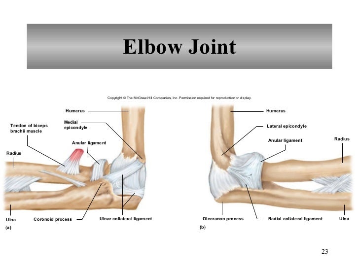
Ligaments of your elbow joint play an essential role in maintaining stability. We'll take a look at those ligaments now. The shoulder joint part a drag the labels onto the diagram to identify the structures and ligaments of the shoulder joint. Ligaments of axis and occipital joint. Injuries may require physical therapy to regain full mobility.
HW 4.pdf - HW 4 Due 11:59pm on Friday October 6 2017 To understand how points are awarded read ... from www.coursehero.com You need to be a group member to play the tournament. No ligaments connect the bones at this joint. Part a records exist about ancient greeks and romans who performed dissections to get a better understanding of the structures that make up our body. Total shoulder movement is made up of the movement from muscles, ligaments, cartilage and other joint structures can be seen with both mri and us. 8 name the arteries and the nerves that coracohumeral ligament : By lack of ligaments, the joint delegates the function of stability fully to the muscles that attach the when the posterior structures of the glenohumeral joint are shortened, this may compromise the in fact, some authors have identified internal impingement as the leading cause of rotator cuff lesions in. The relative degrees of stability and mobility are a reflection of shoulder complex movements represent care fully orchestrated motion of all of its components. Joints of shoulder region at cram.com.
Flexion of the shoulder joint occurs when the humerus (upper arm) moves forwards from the rest of the body, which happens at the end of an underarm throw or bowl in rounders.
The main organs of the body have ordinary english names and doctors use these words. Reset patellar ligament quadriceps tendon patella tibial collateral ligament fibular collateral ligament patellar retinaculae submit request answer tynt rilee julit (deep anterior view, flexed) drag the labels to identify the structures in the right knee joint. Ligaments of axis and occipital joint. But when an adjective is needed they often use an anatomical word. 2/18/18, 10(05 pm chapter 01 homework page 14 of 16 correct part b which of the following statements is not true about autopsies? Join group, and play just play. Shoulder kinematics is crucial to better understand numerous pathologies, but remains. Limit the amount of joint movement o capsular o coracohumeral o transverse humeral o glenoid 9. There are two ways to categorize joints. Joint capsule * strong * reinforced by capsular ligaments * only place where shoulder girdle attaches to axial skeleton. Articulations between the trochlea of the humerus with the ulna and the capitulum of the humerus with the head of the radius comprise the joint. Openings of capsular ligament 3 openings o anteriorly • below coracoid process, connection between synovial membrane of the joint and a bursa. Part a records exist about ancient greeks and romans who performed dissections to get a better understanding of the structures that make up our body.
Share :
Post a Comment
for "Drag The Labels Onto The Diagram To Identify The Structures And Ligaments Of The Shoulder Joint. : Skeletal System Anatomy and Physiology | Nursing School and Study Guides | Human anatomy ..."


Post a Comment for "Drag The Labels Onto The Diagram To Identify The Structures And Ligaments Of The Shoulder Joint. : Skeletal System Anatomy and Physiology | Nursing School and Study Guides | Human anatomy ..."